
Anthony G. Christodoulou, Haosen Zhang, Bo Zhao, T. Kevin Hitchens, Chien Ho, and Zhi-Pei Liang
Volume: 60, Issue: 11, Page(s): 3083-3092

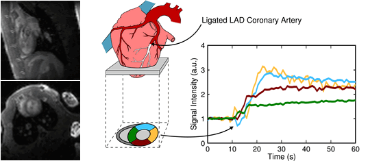
Magnetic resonance imaging (MRI) has long been recognized as a powerful tool for cardiovascular imaging because of its unique potential to measure blood flow, cardiac wall motion and tissue properties jointly. However, many clinical applications of cardiac MRI have been limited by low imaging speed. In this paper, we present a novel method to accelerate cardiovascular MRI through the integration of parallel imaging, low-rank modeling, and sparse modeling. This method consists of a novel image model and specialized data acquisition. Of particular novelty is the proposed low-rank model component, which is specially adapted to the particular low-rank structure of cardiovascular signals. Simulations and in vivo experiments were performed to evaluate the method, as well as an analysis of the low-rank structure of a numerical cardiovascular phantom. Cardiac imaging experiments were carried out on both human and rat subjects without the use of ECG or respiratory gating and without breath holds. The proposed method reconstructed 2-D human cardiac images up to 22 fps and 1.0 mm × 1.0 mm spatial resolution, even in patients with cardiac arrhythmias. After ligating the left anterior descenting (LAD) coronary artery in rats, 3-D cardiac imaging experiments at 67 fps and 0.65 mm × 0.65 mm × 0.31 mm spatial resolution identified regions of hypoperfusion associated with LAD blood supply. The capabilities provided by our method enhance the practical utility of cardiovascular MRI.