
Mihaela Pop, Venkat Ramanan, Franklin Yang, Li Zhang, Susan Newbigging, Nilesh Ghugre, Graham Wright, Sunnybrook Research Institute, University of Toronto
Volume 61, Issue 12, Page: 2930-2938

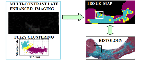
Magnetic resonance imaging (MRI) has exceptional soft tissue contrast and can non-invasively provide critical information to improve the management of patients with prior myocardial infarct. The substrate of potentially lethal cardiac arrhythmias in these patients often resides in the gray zone (GZ), a mixture of viable myocytes and fibrotic collagen strands (replacing dead myocytes) found between healthy myocardium and infarct core (IC). Our efforts are focused on developing methods that can identify such subtle structural heterogeneities as areas of intermediate T1 relaxation in gadolinium-enhanced MRI of the heart. Specifically, in this work, we aimed to demonstrate the correspondence between regions delineated in T1* (apparent T1) maps and tissue characteristics seen in histopathology, and to determine the MR imaging resolution needed to adequately identify GZ-associated substrate. For this, a novel 3D multi-contrast late enhancement (MCLE) MR method was developed and used to image ex vivo swine hearts with chronic infarction at high resolution (0.6×0.6×1.25 mm). Pixel-wise classified tissue maps were calculated using steady-state and T1* images as input to a fuzzy-clustering algorithm. Quantitative histopathological analysis (based on collagen-specific stains) showed very good correlations between histologically-determined areas of heterogeneous and dense collagenous fibrosis, and the corresponding GZ and IC in tissue classified maps. Additionally, we performed volumetric measurements of GZ and IC at high and low image resolutions, and found that the IC volume remained relatively unchanged across all resolutions, whereas the GZ volume progressively increased with diminished image resolution. The results suggested a 1x1x2.5mm3 resolution may be sufficient to adequately identify the GZ from MCLE images, enabling an effective MR probing of remodelled myocardium in chronic infarct. Future work will focus on translating these findings to optimizing the current in vivo MCLE imaging of the GZ.
Keywords: cardiac MRI, image analysis, fibrosis, quantitative T1 maps, gray zone