

Kai Yu, Abbas Sohrabpour, Bin He, University of Minnesota, USA
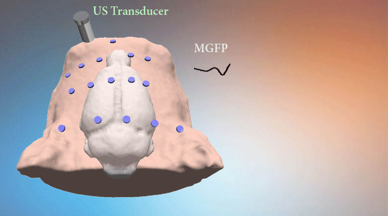
Transcranial focused ultrasound (tFUS) has been introduced as a noninvasive neuromodulation technique with good spatial selectivity. However, there is an unanswered question of whether low-intensity tFUS (much lower than FDA’s regulations) can induce EEG responses or not. There is also an unmet need to develop a non-invasive approach that is able to localize and indicate the brain regions activated by the low-intensity ultrasound in vivo. We report an experimental investigation to noninvasively detect electrophysiological response induced by low-intensity tFUS in an in vivo animal model, and perform electrophysiological source imaging (ESI) of tFUS-induced brain activity from noninvasive scalp EEG recordings. A single ultrasound transducer with customized ultrasound collimator was used to generate bursts of low-intensity ultrasound pulses (spatial-peak temporal-average intensity: Ispta<1 mW/cm2) and induce brain activation at multiple selected sites in an in vivo rat model. Up to 16 scalp electrodes were used to record tFUS-induced EEG. The sonicating event related potentials (ERPs) were analyzed in time, frequency, and spatial domains. Current source distributions were estimated by ESI to reconstruct spatio-temporal distributions of brain activation induced by tFUS. Neuronal activation was observed following low-intensity tFUS, for various tFUS intensities and sonication durations. ESI revealed initial focal activation in cortical area which was induced by the tFUS, and the propagation of activity to surrounding areas over time. The present results demonstrate the feasibility of noninvasively recording brain electrophysiological response in vivo following low-intensity tFUS stimulation, and the feasibility of imaging spatio-temporal distributions of brain activation as induced by tFUS in vivo. The present approach may lead to a new means of imaging brain activity using tFUS perturbations as well as a closed-loop ESI-guided tFUS neuromodulation modality. Website: http://helab.umn.edu/
Keywords: EEG source imaging, In vivo animal model, Perturbation-based neuroimaging, Transcranial focused ultrasound (tFUS)