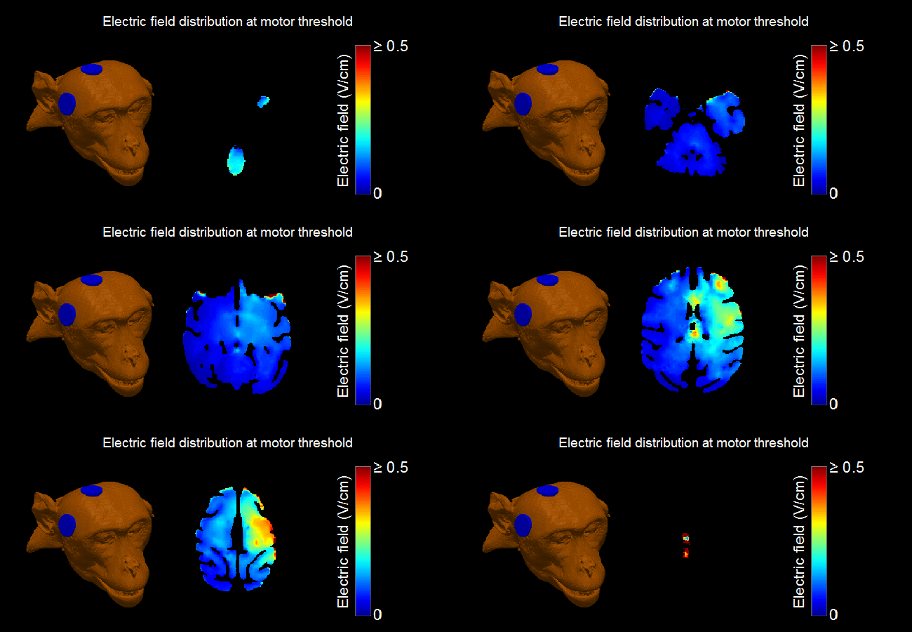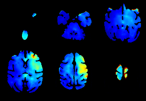
This paper describes a pipeline for realistic head models of nonhuman primates for simulations of noninvasive brain stimulation, and uses these models together with empirical threshold measurements to demonstrate that the models capture individual anatomical variability. Based on structural magnetic resonance imaging data, we created models of the electric field induced by right unilateral electroconvulsive therapy (ECT) in four rhesus macaques. The individual motor threshold was measured with transcranial electric stimulation administered through the ECT electrodes in the same subjects. The interindividual anatomical differences resulted in 57% variation in the median electric field strength in the brain at fixed stimulus current amplitude. Individualization of the stimulus current by the motor threshold reduced the electric field variation in the target motor area by 27%. There was a significant correlation between the measured motor threshold and the ratio of simulated electrode current and electric field strength. Exploratory analysis revealed significant correlations of this ratio with anatomical parameters including the superior electrode-to-cortex distance, vertex-to-cortex distance, and brain volume. The neural activation threshold was estimated to be 0.45 ± 0.07 V/cm for 0.2 ms stimulus pulse width. In conclusion, these results suggest that our individual-specific models of the electric field induced in the brain appropriately capture individual anatomical variability relevant to the dosing of various forms of transcranial electric stimulation including ECT. While these findings are exploratory due to the small number of subjects, this work can contribute insight in nonhuman primate studies of ECT and other brain stimulation interventions, help link the results to clinical studies, and ultimately lead to more rational brain stimulation dosing paradigms.

