
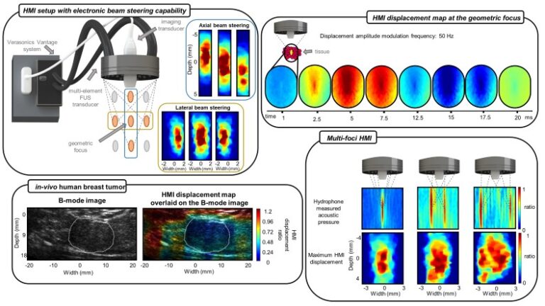
Harmonic motion imaging (HMI) is an ultrasound-based elasticity imaging technique that assesses the mechanical properties of tissues by inducing harmonic displacements. In conventional HMI, a focused ultrasound (FUS) transducer generates the harmonic displacements, and an imaging transducer acquires channel data for displacement estimation, where each transducer is driven using a separate ultrasound data acquisition (DAQ) system. However, the localized and fixed position of the FUS focal spot limits the imaging area and depth of tissue of interest. Additionally, it requires mechanical translation of the transducers, which is challenging and prolongs the imaging time. This study focuses on developing and characterizing a new clinical HMI setup for faster and more convenient data acquisition.
A multi-element FUS transducer with electronic beam steering of ±5 mm in the axial and ±2 mm in the lateral direction from the geometric focus generated the oscillatory force. A pulse sequence was developed to integrate the tracking and excitation pulses into a single signal, driving both transducers synchronously using a single DAQ system. The setup’s performance was validated through hydrophone measurements, experiments on a tissue-mimicking phantom with embedded inclusions, and an in-vivo human breast tumor. Lastly, the FUS was programmed to simultaneously generate multiple foci, enlarging the engaged area.
In-silico results show that the combination of electronic beam steering with the mechanical translation of the HMI transducers accelerated the data acquisition by a factor of 4.5-5.2 compared to the sole mechanical translation of the transducers, without compromising the contrast-to-noise ratio (CNR). Furthermore, applying the combined imaging technique successfully differentiated the in-vivo tumor from the perilesional tissues while reducing the imaging time by about 3.5 times. The new and compact HMI system allows for faster and more flexible data collection and can enlarge the engaged area, which are critical for the imaging and treatment of breast cancer.