

Ahsan Javed, Yoon-Chul Kim, Michael C.K. Khoo, Sally L. Davidson Ward, and Krishna S. Nayak, University of Southern California, USA, Volume 63, Issue 2, Pages:431-437
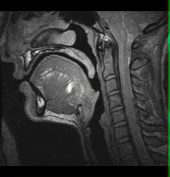
Obstructive sleep apnea is a common breathing disorder in which airflow is blocked during sleep due to collapse of the airway. It is a significant public health issue affecting 4-9% of adults and 2% of children. Our lab has recently developed 3D dynamic MRI technology to non-invasively study the upper airway during natural sleep with high spatio-temporal resolution, and to evaluate patterns of airway obstruction.
When used to capture natural collapses, these scans are approximately 20 min long and contain 1200-2000 frames. Automated segmentation and collapse detection tools are a major unmet need. Manual segmentation of these data requires 10 minutes per frame and is therefore not practical. Visual inspection of each frame is similarly time consuming and not practical. This paper describes a new tool that provides semi-automated identification of time periods of airway obstruction, visualization of collapse sites, and volumetric analysis.
Our approach uses 3D-region growing to segment the air space in 3D dynamic MRI data of pharyngeal airway. This requires 2 minutes of operator time per data set and 5 sec per frame of CPU time. Our method compares well with manual segmentation, resulting in Dice Coefficients of 0.84 to 0.94. For reference, the intra-observer variability of manual segmentation is 0.89 to 0.97. The new algorithm is able to automatically detect 83% of collapse events.