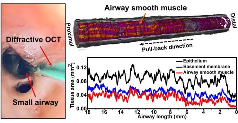
At present, the resolution and contrast of conventional endoscopic optical coherence tomography (OCT) operating at 1300 nm remain challenging to directly visualize the fine microstructures of small airways in vivo. In addition, the increasing volume of OCT images and the shortage of available human expertise impose great challenges on manual image interpretation. To overcome the obstacle, we developed a deep-learning assisted diffractive OCT technology to noninvasively and quantitatively imaging subsurface microcompartments in small airways in three dimensions (3D).
We adopted our recently developed 800-nm diffractive OCT technology with a resolving power of 1.7 µm (in tissue) and improved image contrast to enable direct delineation of airway microstructures. We further customed a ResNet based method to automatically segment, quantify and visualize 3D microcompartments in small airways. The deep-learning based automated method provided an accuracy similar to our experienced investigators (with an average IoU of about 0.92) for segmenting microcompartments in small airways. The feasibility of deep-learning assisted diffractive OCT technology was first demonstrated to directly visualize and quantify critical tissue compartments of small airways in sheep.
Given the high resolution and high segmentation accuracy, deep-learning assisted diffractive OCT demonstrates the ability to quantify and visualize the 3D tissue microcompartments, such as airway smooth muscle, in small airways, thereby enabling the in vivo objective assessment of airway pathology in ways there were previously not possible, allowing a better understanding and management of obstructive lung disease, such as asthma and chronic obstructive pulmonary disease (COPD).

