
Mitko Veta, Josien P.W. Pluim, Paul J. van Diest, and Max A. Viergever
University Medical Center Utrecht, Utrecht, The Netherlands, Volume 61, Issue 5, Page: 1400-1411
Pathology labs are currently undergoing a transformation towards a fully digital workflow. In addition to the digital management of tissue samples, pathology orders and reports, this includes the digitization of histopathology slides, which aims to replace the optical microscope as the primary tool used by pathologists. This transformation has only recently been enabled by the introduction of cost and time efficient whole slide imaging (WSI) scanners.
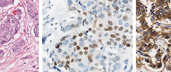
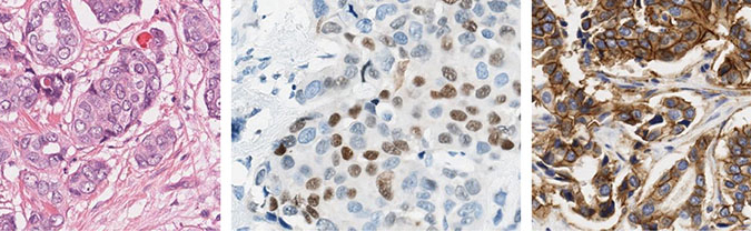
A relatively large percentage of the samples that are analyzed in pathology labs are from breast cancer patients, since this disease is the most prevalent form of cancer among women. Analysis methods that are routinely performed by pathologists, such as determination of the histological grade and the hormone receptor status by immunohistochemistry, can be tedious and are hampered by observer variability. The histological tumor grade is commonly determined according to the modified Bloom-Richardson system, which consists of semi-quantitative assessment of nuclear atypia, tubule formation and mitotic activity. The analysis of immunohistochemically stained slides mainly involves the estimation of the number of cells that are positive for a particular antigen and the degree of positivity (staining intensity).
One of the main benefits of digital slides compared to conventional glass slides is that they enable the use of quantitative automatic image analysis methods. These methods have the potential to tackle the problems that stem from the subjective interpretation by pathologists and, at the same time, reduce their workload.
In this paper, we give an overview of image analysis methods that have been proposed for breast cancer histopathology images. We focus on automatic image analysis of histopathology tissue preparations imaged by brightfield microscopy, since this covers the bulk of the work that is performed by pathologists for this disease.