

Kristen L. Lurie, Roland Angst, and Audrey K. Ellerbee, Stanford University
Volume 61, Issue 7, Page: 2141-2153
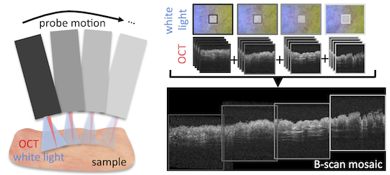
We demonstrate the first automated, volumetric mosaicing algorithm for optical coherence tomography (OCT) that both accommodates 6-DOF rigid transformations and implements a bundle adjustment step amenable to generating large fields of view with endoscopic and freehand imaging systems. Our mosaicing algorithm exploits the known, rigid connection between a combined white light and OCT imaging system to reduce the computational complexity of traditional volumetric mosaicing pipelines. Specifically, the search for 3D point correspondences is replaced by two, 2D processing steps: we first co-register a pair of white light images in 2D and then generate a surface map based on the volumetric OCT data, which is used to convert 2D image homographies into 3D volumetric transformations. A significant benefit of our dual-modality approach is its tolerance for feature-poor datasets such as bladder tissue; in contrast, approaches to mosaic feature-rich volumes with significant variations in the local intensity gradient (e.g., retinal data containing prolific vasculature) are not suitable for such feature-poor datasets. We demonstrate the performance of our algorithm using ex vivo bladder tissue and a custom tissue-mimicking phantom. The algorithm shows excellent performance over the range of volume-to-volume transformations expected during endoscopic examination and comparable accuracy with and several orders of magnitude superior run times than an open-source gold-standard algorithm (N-SIFT). We anticipate the proposed algorithm can benefit bladder surveillance and surgical planning. Furthermore, its generality gives it broad applicability and potential to extend the use of OCT to clinical applications relevant to large organs typically imaged with free-hand, forward-viewing endoscopes.
Research website: http://sbo.stanford.edu/