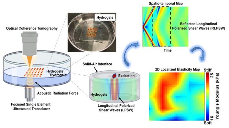
Scaffolds play an important role in tissue engineering because they provide a supporting structure to mimic a native extracellular matrix (ECM) micro-environment, thereby allowing for cell adhesion, proliferation, and migration. By designing various elastic properties of hydrogels, living cells can be combined with the scaffolds to engineer living hydrogels (cell-laden scaffolds). Different requirements exist for hydrogel scaffolds for each type of engineered tissues. For example, compositions and cross-linked collagen densities in hydrogels significantly affect their mechanical properties, which affect the functionality of growing tissues with respect to cell migrations and differentiations. Also, softer scaffolds provide a higher cell proliferation than stiffer scaffolds. Therefore, exploring elastic properties and temporal evolution of hydrogel properties is critical in tissue engineering and hydrogel mechanics. A standard method to determine elastic properties of hydrogels relies on dynamic mechanical analysis (DMA). However, drawbacks of DMA are that is a destructive measurement and provides a global elastic value which may hamper the evaluation in biological living hydrogels and various heterogeneous materials. In this paper, we report a new elastography modality named acoustic force elastography microscopy (AFEM), which has ability to resolve localized and two-dimensional (2D) elastic properties of both transparent and opaque materials such as hydrogels or biological thin tissues with advantages of being non-contact and non-destructive. The phenomenon of reflected longitudinally polarized shear waves (RLPSWs) was utilized as the theoretical foundation of AFEM. In future applications, AFEM will be used to explore the continuous evolution of properties of engineered living soft materials associated with various situations, such as tissue growth, cell proliferation, cancer organoids or hydrogel degradation in tissue engineering, material science and precision medicine applications.

Acoustic Force Elastography Microscopy
Sign-in or become an IEEE member to discover the full contents of the paper.
