The 21st century has embraced miniaturization and witnessed the emergence of multidisciplinary nanotechnology in the medical field, popularly known as nanomedicine. It has long been established that chronic infections such as hepatitis and malaria, as well as diseases such as cancer and arthritis, are diverse, complex, and heterogeneous, wherein patients do not respond identically to the same spectrum of drugs. Traditional bolus delivery of biotherapeutics offers medical practitioners limited control over dosage, tissue distribution, and bioavailability in vivo. Many therapeutic agents, particularly chemotherapy drugs such as Paclitaxel, are highly toxic to the body, and therefore, limited dosage can be administered in a patient. Similarly, soluble protein growth factors have limited potential to penetrate cell membrane barriers to transform the functioning and fate of cells. Consequently, the effective delivery of biotherapeutics is a major goal of current biomaterial-based strategies.
As material scientists and chemists develop new classes of biomaterials, bioengineers and clinicians are finding diverse applications for these materials in medicine. Myriad biomaterials have been engineered into nanoscale constructs for use as therapeutics against cancer, as arthritis treatments, or for manipulating the behavior of cells. Nanomaterials offer the ability to better target specific areas in the body and deliver agents with better-controlled timing, delivery rate, and dosage. The nanobiomaterials available today are engineered polymeric nanoparticles, DNA gels, nanoshells, lipid-based platforms, dendrimers, viral constructs, and magnetic nanoparticles, with a continuous influx of novel nanotechnology platforms emerging at an increasing rate. Already, there are several nanoengineered particle–drug combinations available on the market and/or in clinical trials.
More than 20 nanoparticle therapeutics have been approved by the U.S. Food and Drug Administration (FDA) for clinical use. Some of the common nanoparticle platforms for therapeutic delivery of drugs and biomolecules include lipid-based liposomes and polymer-based particles such as methoxy-polyethylene glycol (PEG)-poly[D,L-lactide]taxol. Doxil is the first drug-encapsulated liposomal formulation for doxorubicin delivery that is on the market and has been approved for the treatment of AIDS-related Kaposi’s sarcoma, ovarian cancer, and multiple myeloma. Doxil, which consists of the drug Doxorubicin packaged inside PEG-modified liposomes was designed to limit exposure of the cardiotoxic drug to the tumor by taking advantage of the enhanced permeability and retention (EPR) effect. With the EPR effect, the concentration of liposomes tends to increase inside solid tumors because of leaky architecture of the blood vessels compared to healthy tissues.
Polymer–drug conjugates represent a newer class of nanotherapeutics that have been used in delivering drugs, proteins, and nucleic acids. Linear polymers such as N-(2-hydroxypropyl)methacrylamide are hydrophilic and nonimmunogenic and currently under clinical trials for treatment of primary hepatocellular carcinoma. Ligands can also be attached to these conjugates to attain cell specificity. Yet, the ultimate potential of nanomedicine can only be realized when we demonstrate the ability to selectively deliver nanobiomaterials to the tissues of interest, with controlled release and degradation rates, and minimal accumulation in nonspecific healthy tissues.
One of the most promising of the recent tools are smart nanoparticles consisting of “signaling” and “receiving” modules that communicate through the cell signaling network within the body. von Maltzahn et al. [1] engineered signaling modules such as PEG-modified gold nanorod and tumor-targeted human protein and receiving modules containing iron oxide nanoworms and therapeutic liposomes. Using photothermal effect, the signaling module initiates blood coagulation cascade and broadcasts the tumor location to the receiving module, which is then recruited using the coagulation cascade. Such communicating nanoparticle systems represent the next generation of autonomous tools, moving toward the improvement of targeted cancer therapy and diagnostics.
One of the most fascinating developments has been in the use of nanomaterials for gene therapy applications such as delivering DNA to cells in the body for altering what protein they produce or
for delivering gene silencing RNA to shut down the production of a particular gene/protein of interest. This way, one can amplify or deplete biological components that interfere with disease treatment or support the growth and development of diseases.
For example, DNA has been used as a vaccine against infections, cancers, and allergies. Delivering DNA itself is challenging because of its short half-life, and the required high doses have so far been insufficient in eliciting significant cellular and antibody response against an infection in humans. The inability to induce significant immune response through naked DNA immunization is partly attributed to physical and molecular barriers such as cellular uptake and inefficient gene transfection. However, complexation of disease-specific DNA with cationic polymers can significantly improve the cellular uptake, preserve DNA integrity, and the immunogenicity of the protein for which the DNA encodes. Cationic polymers such as poly(l-lysine) [2] and polyethylenimine [3], [4] have been extensively used for DNA delivery for preclinical immunization purposes. Additionally, seminal work by Roy et al. has demonstrated the effectiveness of engineered chitosan–DNA nanoparticles in inducing protective immunity in the peanut allergy mouse model through oral delivery [5].
The use of nanoparticles in vaccines continues to grow, including an important study that shows a pathogen-mimicking nanoparticle made of poly(lactic-co-glycolic) acid (PLGA), an FDA-approved polymer, carrying a select combination of immune-specific drugs induced robust immunity against the 2009 pandemic H1N1 influenza A in mice and nonhuman primates [6]. The power of nanotechnology with combined chemistry has been demonstrated by Reddy et al., where Pluronic-stabilized polypropylene sulfide nanoparticles of 25-nm size successfully exploited the interstitial body fluid transport compared to 100-nm particles to accumulate in the lymph nodes—a primary site for immune reactions. Interestingly, nanoparticles with hydroxyl groups in the surface activated the immune complement system in mouse models compared to methoxylated groups.
A unique application of engineered therapeutic nanoparticles is in the delivery and enhanced retention of anti-inflammatory biomolecules in the knees of patients with osteoarthritis. Osteoarthritis is a common joint disorder where the repair of cartilage involves sequential interplay of growth factors with potential knee (synovial) inflammation by a very specific set of proteins called inflammatory cytokines. Anti-inflammatory mediators, such as the Interleukin-1 receptor antagonist (IL-1Ra), reduce inflammation associated with arthritis, bone resorption, and pain levels, and, at high local concentrations, slow down osteoarthritis progression in animal models.
Knee or intra-articular delivery of therapeutics to modulate such damaging proteins in osteoarthritis is challenging and bolus protein injections of IL-1Ra suffer from rapid clearance and reduced potency over time. We recently reported self-assembling nanoparticles presenting IL-1Ra for enhanced delivery, retention, and bioactivity in the rat stifle joint (analogous to the knee) [7]. Using block copolymers that self-assembled into 300-nm-diameter particles and efficiently bound IL-1Ra, we demonstrated specific targeting of cells residing in joints (synoviocyte) via surface IL-1 receptors and inhibition of IL-1-dependent signaling cascades. These nanoparticles demonstrated a significantly longer retention time of IL-1Ra in the rat stifle joint than soluble IL-1Ra (Figure 1) and no alterations in knee cartilage structure and morphology in healthy animals. Compared to solid hydrophobic polymeric nanoparticles, such as PLGA, our nanoparticles provide a hydrophilic environment that would maintain protein structure and bioactivity compared to hydrophobic surfaces. In addition, self-assembling nanoparticles do not require harsh solvent conditions that often reduce the bioactivity of drug or biomolecules. Finally, unlike most existing nanoparticles, our nanoparticles incorporate a protein tethering moiety on the surface and do not require reengineering the biomaterial to conjugate target–cell specific ligands.
![FIGURE 1 (a) Anti-inflamatory IL-1Ra-tethered nanoparticles (inset: scanning electron micrograph) are distributed throughout the intra-articular joint space in a rat knee. IL-1Ra was tagged with a Dylight-IR-650 dye before tethering IL-1Ra to particles for visualization purposes. (b) Soluble protein is rapidly cleared from the injection site, indicating poor retention in a rat knee. (Figure adapted with permission from [7].)](https://www.embs.org/wp-content/uploads/2014/03/32221.png)
A recent paradigm shift in nanomedicine has been toward fabricating shape-specific nanoparticles for therapeutic delivery and diagnostics. Inspired from shapes of microbes and pathogens in nature, several research groups have generated nanofabrication techniques such as particle replication in nonwetting templates (PRINT) and jet and flash imprint lithography (J-FIL) to generate nanoparticles with precisely controlled shape and geometry. Using the J-FIL technology, Agarwal et al. [8] demonstrated the differential dependence of nanoparticle shape on uptake by the cells. Disc-shaped nanogels made of PEG polymer were internalized more efficiently than nanorods by immune cells as well as epithelial and endolthelial cells. The importance of nanoparticle shape has been further implicated in tumor targeting studies by Chauhan et al. [9], where the group demonstrated enhanced penetration of nanorods over spherical-shaped nanoparticles due to improved transport properties. Therefore, design considerations that take into account bulk material property, size, shape, and surface chemistry could be critical for directing therapies against tumor, infections, as well as retention and penetration within tissues.
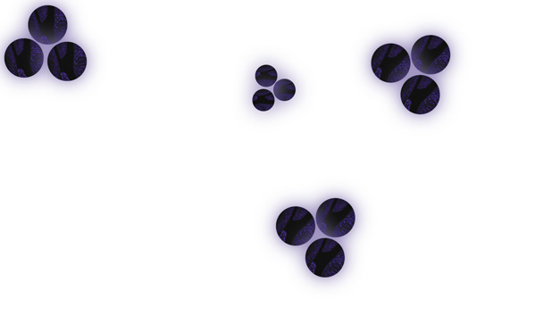
The use of nanomedicine in engineering cells is a relatively new area that aims to procure advances in cell-based therapies, such as immune and stem cells. Nanoscale engineering further provides innovative tools to manipulate elements of cell and tissue engineering that have been previously difficult to control. Several types of bioactive nanomaterial that have clinical relevance have been reported for musculoskeletal tissue engineering in the last few years, including bioactive glasses, tricalcium phosphate, calcium sulfate, hydroxyapatite, and glass–ceramics. Difficulties persisting with many of these known materials include lack of intrinsic properties, poor processing abilities that would induce and enhance bonelike transformations (osteoinduction), and insufficient degradation of nanoparticles. Gaharwar et al. [10] reported bioactive disc-shaped nanoparticles based on synthetic silicate (Figure 2), which is nontoxic to cells and promotes bonelike differentiation of human mesenchymal stem cells in the absence of any growth factor such as bone morphogenetic proteins-2 or dexamethasone. These synthetic silicates are made up of simple or complex salts of silicic acids, magnesium, and sodium, which can be easily degraded under in vivo conditions. Such bioactive nanodiscs that resemble platelets shapewise may be used to develop devices such as injectable matrixes, therapeutic agents, and bioactive fillers for stimulating specific cellular responses in musculoskeletal-related tissue engineering.
![FIGURE 2 Disc-shaped silicate nanoplatelets induce osteogenic differentiation of human mesenchymal stem cells in the absence of any osteoinductive factor. (Figure adapted with permission from [10].)](https://www.embs.org/wp-content/uploads/2014/03/32273.png)
Conclusion and Perspective
Advances in nanobiotechnology and material science have resulted in a diverse toolkit of nanosystems with unique physiochemical properties. Nanobiomaterials-based drug delivery, diagnostics, and cell engineering approaches will continue to see rapid expansion over the next several decades. While the potential is clear, there are a few challenges. Problems that plague several existing polymer nanoparticles include inherent polymer toxicity, stability of the polymer–drug linkage, and the immunogenic potential of the polymer. For example, natural polysaccharide dextran conjugated doxorubicin (AD-70) resulted in severe hepatotoxicity and thrombocytopenia in the phase 1 trial. Additionally, methods to control particle shape are few and expensive with little flexibility with choice of biomaterial. Therefore, continued research efforts are needed to engineer nanoscale systems that not only deliver therapeutics or manipulate the behavior of a cell but also to do this is in a systematically coherent manner. As these issues are explored, we will see new cures and treatments emerge.
References
- G. von Maltzahn, J. H. Park, K. Y. Lin, N. Singh, C. Schwoppe, R. Mesters, W. E. Berdel, E. Ruoslahti, M. J. Sailor, and S. N. Bhatia, “Nanoparticles that communicate in vivo to amplify tumour targeting,” Nature Mater., vol. 10, no. 7, pp. 545–552, July 2011.
- E. Wagner, M. Ogris, and W. Zauner, “Polylysine-based transfection systems utilizing receptor-mediated delivery,” Adv. Drug Deliv. Rev., vol. 30, nos. 1–3, pp. 97–113, Mar. 1998.
- D. Nguyen, J. Green, M. Chan, R. Langer, and D. Anderson, “Polymeric materials for gene delivery and DNA vaccination,” Adv. Mater., vol. 20, pp. 1–21, 2008.
- K. Regnstrom, E. G. Ragnarsson, M. Koping-Hoggard, E. Torstensson, H. Nyblom, and P. Artursson, “PEI—A potent, but not harmless, mucosal immuno-stimulator of mixed T-helper cell response and FasL-mediated cell death in mice,” Gene Ther., vol. 10, no. 18, pp. 1575–1583, Sep. 2003.
- K. Roy, H. Q. Mao, S. K. Huang, and K. W. Leong, “Oral gene delivery with chitosan–DNA nanoparticles generates immunologic protection in a murine model of peanut allergy,” Nature Med., vol. 5, no. 4, pp. 387–391, Apr. 1999.
- S. P. Kasturi, I. Skountzou, R. A. Albrecht, D. Koutsonanos, T. Hua, H. I. Nakaya, R. Ravindran, S. Stewart, M. Alam, M. Kwissa, F. Villinger, N. Murthy, J. Steel, J. Jacob, R. J. Hogan, A. Garcia-Sastre, R. Compans, and B. Pulendran, “Programming the magnitude and persistence of antibody responses with innate immunity,” Nature, vol. 470, no. 7335, pp. 543–547, Feb. 2011.
- R. E. Whitmire, D. S. Wilson, A. Singh, M. E. Levenston, N. Murthy, and A. J. Garcia, “Self-assembling nanoparticles for intra-articular delivery of anti-inflammatory proteins,” Biomaterials, vol. 33, no. 30, pp. 7665–7675, Oct. 2012.
- R. Agarwal, V. Singh, P. Jurney, L. Shi, S. V. Sreenivasan, and K. Roy, “Mammalian cells preferentially internalize hydrogel nanodiscs over nanorods and use shape-specific uptake mechanisms,” Proc. Natl. Acad. Sci. U.S.A., Oct. 2013.
- V. P. Chauhan, Z. Popovic, O. Chen, J. Cui, D. Fukumura, M. G. Bawendi, and R. K. Jain, “Fluorescent nanorods and nanospheres for real-time in vivo probing of nanoparticle shape-dependent tumor penetration,” Angew. Chem., vol. 50, no. 48, pp. 11417–11420, Nov. 2011.
- A. K. Gaharwar, S. M. Mihaila, A. Swami, A. Patel, S. Sant, R. L. Reis, A. P. Marques, M. E. Gomes, and A. Khademhosseini, “Bioactive silicate nanoplatelets for osteogenic differentiation of human mesenchymal stem cells,” Adv. Mater., vol. 25, no. 24, pp. 3329–3336, June 2013.