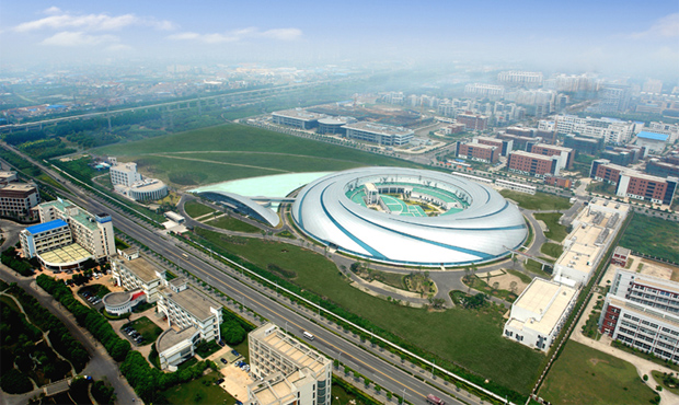
Above: The Shanghai Synchrotron Radiation Facility (SSRF).
Since its development in 1947, synchrotron radiation (SR) X-rays have had tremendous applications in many fields, including in physics, material science, the life sciences, and medicine. The bright X-ray beam produced by synchrotron allows for the study of structures at an atomic level and events that occur at literally the speed of light. Initially, SR medical imaging was used to detect the detailed structures of various cancers in the breast, lung, and liver, as well as cerebrovasculature and cardiovasculature ex vivo. In more recent years, several third generation SR facilities have used synchrotron radiation in angiography on live animals, opening a new area of study on the effects of blood flow and microvascular function in vivo in many organ systems. A major advantage is that SR X-rays subject patients to potentially less radiation exposure than conventional X-ray imaging. This article introduces the applications of SR biomedical imaging in small animal disease models at beam line BL13W in the Shanghai Synchrotron Radiation Facility (SSRF) in China. We summarize the studies of rodent cerebral vasculature under ischemic or hemorrhagic conditions and the effort in developing absorption-based and phase-sensitive tumor angiography techniques aimed at early detection of micro-vessel growth.
Charged particles emit light when they are accelerated. If electrons are constrained to move in a curved path, they will accelerate toward the inside of the curve and will also radiate what is called synchrotron light. Light source facilities use particle accelerators to generate super-bright, intense beams of electromagnetic radiation. This radiation is referred to as light. The resulting light spectrum is very broad, including invisible X-rays, ultraviolet light, and infrared light.
Accelerator-based synchrotron light was first observed at the General Electric Research Laboratory in the United States in 1947. In the 1960s, it began to be regarded as light with exceptional properties. The light produced from today’s light sources is very bright. In other words, the beam of X-rays or other wavelengths is as thin as a hair and very intense. Just as laser light is much more intense and concentrated than the beam of light generated by a flashlight, so an x-ray beam produced by a synchrotron is a trillion times brighter than the beam produced by a hospital x-ray machine whose x-ray tubes emit light over a wide angle and with high intensity only at particular wavelengths [1].
Currently, the SSRF biomedical imaging beam line mainly serves for pre-clinical animal studies for models of various diseases. At the European Synchrotron Radiation Facility in Grenoble, France, studies have been performed on 51 volunteer patients for trial studies of coronary arteries angiography.
Introduction to the Shanghai Synchrotron Radiation Facility (SSRF)
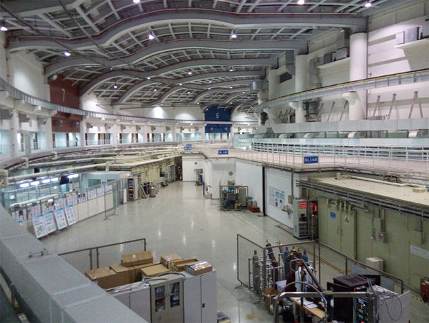
SSRF is a third-generation SR light source. SSRF consists of a 150 MeV linear electron accelerator as injector, a booster that can increase the electron energy from 150 MeV to 3.5 GeV in 0.5 second, and a 3.5 GeV electron storage ring with 432 meter circumference. SR emits light with high flux, high brilliance, and wide photon energy range. There are 40 bending magnets, 16 standard straight sections (6.5 meters), and 4 long straight sections (12 meters) along the ring, thus around 50 beam lines could be constructed around the ring. The initial 7 beam lines, which include STXM-BL08U1A, X-ray imaging-BL13W, XAFS-BL14W, diffraction-BL14B, X-ray microprobe-BL15U, SAXS-BL16B, macromolecular crystallography-BL17U, and one branch beamline-BL08U1B X-ray interference lithography, were formally opened to users on May 6, 2009 [1].
BL13W X-ray Imaging Beam Line
The X-ray imaging beam line takes a 2.0 Tesla hybrid-type wiggler as light source, which is composed of 16 poles and the periodic length is 14 cm, the maximum K-value is 24.8 at minimum gap of 17 mm. A specially designed graphite filter system is utilized to absorb low-energy photons and reduce the heat-load on downstream components such as beryllium window and monochromator. The fixed-exit double crystal monochromator cooled with liquid nitrogen is placed at 28 m away from the source point. Two sets of crystals Si (111) and Si (311) could be interchanged to cover photon energy ranges from 8 KeV to 38 KeV and 38 KeV to 72 KeV, respectively. The beam size is 45 mm (Horizontal)×5 mm (Vertical) at 20 keV.
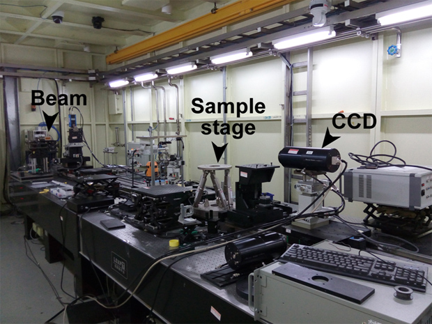
The SR based X-ray allows biomedical imaging to achieve high contrast, high spatial resolution, and high temporal resolution, as a result of the high intensity, high coherence, quasi-parallel and monochromatic properties of the SR beam. The BL13W at SSRF routinely supports absorption-based imaging, phase-sensitive imaging, microCT (µCT) imaging, and X-ray fluorescence imaging. SR-µCT is a non-destructive technique widely used for visualizing the morphology of samples, and for assessing quantitative information on their three-dimensional geometries and properties, which has found extensive applications in different research fields. The data processing and analysis is critical for SR-µCT experiments. Data processing software PITRE, multi-scale 3D structure characterization models, and a pseudo-global tomography algorithm have been developed at the beam line to improve image quality and computation at efficiency.
SR based X-ray fluorescence CT (XFCT), a complement to transmission based CT, is a stimulated emission tomography modality, which allows to reconstruct elemental distributions on a virtual section across the sample by various algorithms. Yang et al. has established an XFCT system at BL13W, and the ordered-subsets expectation maximization algorithm was introduced into XFCT to speed up the data acquisition and improve the image reconstruction [2].
Applications of SR Angiography in a Rodent Stroke Model
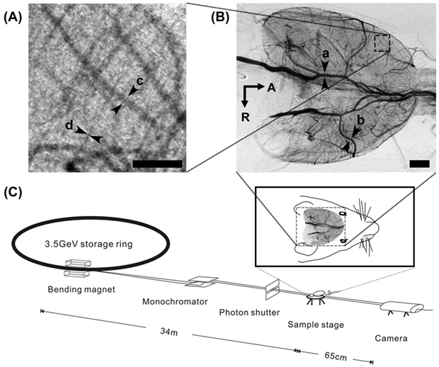
Cerebral vascular disease is a major cause of death in the world, especially ischemic stroke and intracranial hemorrhage (ICH). SR angiography of rodent cerebral vasculature was first developed by Dr. Kidoguchi and his colleagues in 2006 in the Japanese synchrotron facility Spring-8, Japan. At SSRF in Shanghai, we first performed SR angiography in living mice and rats to study the morphology and functions of intracranial vasculatures in cerebral ischemia, intracranial hemorrhage (ICH), and subarachnoid hemorrhage (SAH) models. Our data showed that SR angiography can be used to detect intracranial arteries with a diameter of less than 50 μm, which is much higher than magnetic resonance angiography (MRA) and in vivo microscopic computed tomography (Micro-CT).
SR Angiography in Cerebral Ischemia
SR angiography provides a unique tool for monitoring hemodynamic changes and microvascular morphology in the rodent stroke model. Using SR angiography, we modified the mouse and rat intraluminal suture middle cerebral artery occlusion (MCAO) model, and for the first time, we monitored the cerebral angiogenesis in vivo in the mouse ischemic stroke model after transgenic treatment [3].
The suture MCAO model is used worldwide to study cerebral ischemia. However, several complications are associated with this model, including high animal mortality, low ischemia/reperfusion rates in transient MCAO model, and inadequate occlusion. We found that the variations of dysplasia in posterior communication arteries (PcomA) existed in C57BL/6 mice, which could affect the blood supply in the posterior cerebral artery (PCA) area, thus resulting in the high mortality and instability of intraluminal suture permanent MCAO model. According to the result of SR angiograph, we proved that the high mortality of MCAO model in C57BL/6 mice was mainly due to the dysplasia in PcomA. According to this observation, we optimized the silicone coating of an 8-0 suture tip, which reduced the model mortality to zero after permanent MCAO in C57BL/6 mice and produced stable brain infarct volume independent of the patency of PcomAs. We also evaluated the influence of suture properties in rat intraluminal MCAO model. The SR angiography results, together with histology data, proved that silicone coating length can affect the cerebral blood flow and brain infarct volume after MCAO [4]. Using SR angiography, we also found that the suture-induced thrombosis is a critical element of unreperfusion . Our results showed that appropriate heparin management provides a useful approach for improving reproducibility of the reperfusion model in mice.
Gene therapy treatment in experimental stroke is an area pursued by many stroke researchers. We found that heterologous Netrin-1 (NT-1) overexpression in the ipsilateral hemisphere after cerebral ischemia in mice can significantly improve local angiogenesis and functional recovery. Using SR angiography we verified the functional angiogenesis induced by NT-1 treatment in vivo for the first time [3]. We also demonstrated that human endothelial progenitor cells (EPC) can improve angiogenesis in the ischemic area in mice (Data not published). We anticipate that SR angiography can become the standard method to detect the angiogenesis in experimental research.
SR Angiography in ICH and SAH
ICH and SAH are two types of hemorrhagic stroke with the highest mortality rate in humans. ICH and SAH are mainly caused by spontaneous rupture of the cerebral small vessels with vascular abnormalities, such as arteriovenous malformations (AVM) and aneurysms. Various approaches have been tested on animal models; however, most of the methods were histological analysis in vitro which cannot review the vascular structure and function information under physiological status. So, monitoring the hemodynamic and structural evaluation of cerebral vasculatures in vivo after ICH and SAH has remained critical but still formidable in experimental small animal research. Using SR angiography, we studied the vasospasm of the anterior communicating artery (AcomA) and middle cerebral artery (MCA) in two different ICH models in rats generated by injecting autologous blood into the cisterna magna and prechiasmatic cistern. We identified that both MCA and AcomA shrunk the most at day 3 after SAH and that the diameter of anterior cerebral artery (ACA) in the prechiasmatic cistern injection rat was smaller than that in the cisterna magna injection group, but not for MCA.
We also assessed the blood flow changes in arteries that supply the cerebral cortex and striatum in mice after ICH by combining SR angiography and Laser Speckle Contrast Imaging (LSCI). The results showed that the blood flow in the cortical branches of MCA was significantly decreased at 20 min after blood injection, while recovered after three days. The blood flow in MCA, PCA, and ACA were not changed three days after ICH according to SR angiographic results. Interestingly, the blood flow of lenticulostriate artery (LSA) was significantly increased compared to control group three days after ICH. According to the result above, we concluded that SR angiography provided a novel technique, which could directly evaluate and monitor the structural and functional changes in cerebral vascular in small animal experimental ICH models.
Despite the many advantages of SR angiography, there still remain a number of disadvantages that need to be overcome in the future. Animals could not survive for a long period after SR angiography under the current setting mainly due to radiation injury resulting from the long imaging time required for live imaging. The ionizing radiation injury of brain tissue by SR X-ray is still unclear. The existing data suggests that the tissue tolerance was much lower under SR X-ray compared to conventional X-ray (Data not published). Last but not least, the quantification of vessels using SR angiograph is as big a challenge as in magnetic resonance angiography (MRA) and computed tomographic (CT) angiography.
Application of SR-µCT for tumor angiogenesis
Tumor angiogenesis, the process of new blood vessels sprouting from pre-existing vessels, plays a vital role in cancer growth and development. In vivo imaging of micro-vessels at early stages is an important step in understanding the developing mechanism, early detection, and evaluation of therapeutic effects for tumor diseases. At the BL13W beam line of SSRF, ex vivo and in vivo micro-angiography using SR combined with a high-resolution system has been developed as a powerful tool to study small vessels of tumor diseases. Liu P. et al. investigated physiological features of whole-body mouse microvasculature using barium sulfate as contrast agents for the first time [5]. The 3D reconstruction of lung cancer angiogenesis with barium was obtained with the spatial resolution of up to ∼ 10 μm, which was much higher than that of the conventional angiography.
As a new imaging method, X-ray phase-contrast imaging (XPCI) can greatly improve the image quality of soft tissues, particularly at the interface of tissues, where the refractive index changes significantly, with potential in clinical applications. At SSRF, the blood vessels of excised livers were clearly shown using synchrotron X-ray in-line phase-contrast imaging by replacing the blood with physiological saline. Tang et al. introduced microbubbles into tissue, and microbubbles produced a significant change in the refractive index and highlighted the lumen of the vessel. This study demonstrated that microbubble-based PCI was a promising method to visualize the mouse renal vasculature and tumor-associated vessels using SR phase contrast imaging at the BL13W beam line of SSRF.
In vivo phase-contrast imaging of blood vessels without a contrast agent is still a big challenge. If the intensity difference could be increased between the vessel wall and other surrounding soft tissues, then in vivo phase-contrast imaging of blood vessels might be achieved. Liu P. et al. developed RGD peptides conjugated magnetite nano cluster probes for high resolution micro-CT imaging of tumor angiogenesis and angiogenic vessels were clearly detected by in vivo micro-CT imaging. It is particularly hopeful to realize in vivo SR phase contrast imaging of blood vessels. Further investigation in the SR phase contrast imaging combined with targeted nanoparticle probes are being performed at the BL13W beam line of SSRF with the aim to visualize vascular diseases in the near future.
Conclusions and Perspectives
SR based X-ray imaging has been well developed and used in research related to cerebrovascular diseases and cancer in BL13W beam line of SSRF. Research has proven that the SR angiography was helpful for studying both ischemic and hemorrhage stroke in living rodent models and in tumor angiography. However, further effort is required in reducing the ionizing radiation injury, decreasing the exposure time, and developing methods for three dimensional vessel density quantification in the future to make the SR angiography more applicable in broader ranges of biomedical imaging. With these developments of SR imaging in SSRF and other SR facilities, we expect to see further applications of SR biomedical imaging in basic and clinical researches, diagnoses, and therapies.
References
- SSRF (2014) Website.
- Yang Q, Deng B, Lv W, Shen F, Chen R, et al. (2012) Fast and accurate X-ray fluorescence computed tomography imaging with the ordered-subsets expectation maximization algorithm. J Synchrotron Radiat 19: 210-215.
- Lu H, Wang Y, He X, Yuan F, Lin X, et al. (2012) Netrin-1 hyperexpression in mouse brain promotes angiogenesis and long-term neurological recovery after transient focal ischemia. Stroke 43: 838-843.
- Guan Y, Wang Y, Yuan F, Lu H, Ren Y, et al. (2012) Effect of suture properties on stability of middle cerebral artery occlusion evaluated by synchrotron radiation angiography. Stroke 43: 888-891.
- Liu P, Sun J, Zhao J, Liu X, Gu X, et al. (2010) Microvascular imaging using synchrotron radiation. J Synchrotron Radiat 17: 517-521.