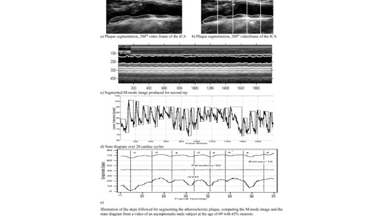
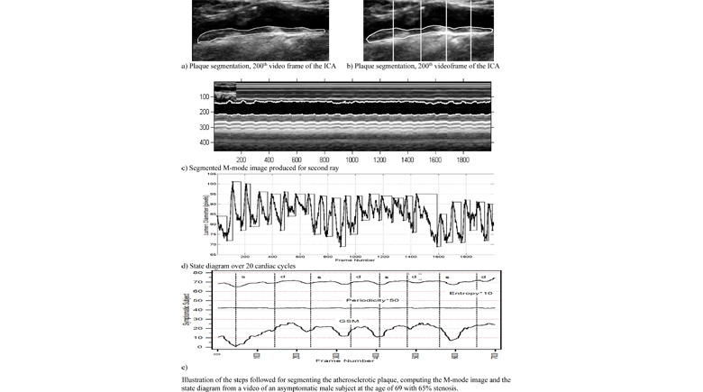
The objective of this paper was to investigate texture feature variability in ultrasound video of the carotid artery during the cardiac cycle in an attempt to define new discriminatory biomarkers of the vulnerable plaque. More specifically, in this paper, 120 longitudinal ultrasound videos, acquired from 40 normal (N) subjects from the common carotid artery and 40 asymptomatic (A) and 40 symptomatic (S) subjects from the proximal internal carotid artery were investigated. The videos were intensity normalized and despeckled, and the intima-media complex (IMC) (from the N subjects) and the atherosclerotic carotid plaques (from the A and S subjects) were segmented from each video, in order to extract the M-mode image, and the texture features associated with cardiac states of systole and diastole. The main results of this paper can be summarized as follows: 1) texture features varied significantly throughout the cardiac cycle with significant differences identified between the cardiac systolic and cardiac diastolic states; 2) gray scale median was significantly higher at cardiac systole versus diastole for the N, A, and S groups investigated; 3) plaque texture features extracted during the cardiac cycle at the systolic and diastolic states were statistically significantly different between A and S subjects (and can thus be used to discriminate between A and S subjects successfully). The combination of systolic and diastolic features yields better performance than those alone. It is anticipated that the proposed system may aid the physician in clinical practice in classifying between N, A, and S subjects using texture features extracted from ultrasound videos of IMC and carotid artery plaque. However, further evaluation has to be carried out with more videos and additional features.