
High-Frequency Oscillations Recorded on the Scalp of Patients with Epilepsy Using Tripolar Concentric Ring Electrodes
https://www.embs.org/jtehm/wp-content/uploads/sites/17/2014/06/feat.jpg
540
508
IEEE Journal of Translational Engineering in Health and Medicine (JTEHM)
//www.embs.org/jtehm/wp-content/uploads/sites/17/2022/06/ieee-jtehm-logo2x.png
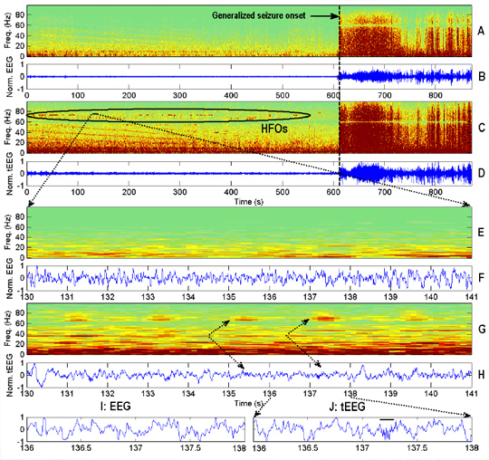
Panel B shows 12 minutes of bipolar EEG from Fp2-F4 (1-70 Hz, 200 S/s). Panel A is the corresponding spectrogram. Panel E shows 30 seconds of EEGfrom Panel B at…
read more