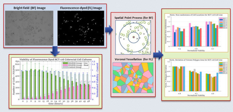
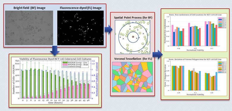
In evaluation of cell viability and apoptosis, spatial heterogeneity is quantified for cancerous cells cultured in 3D in-vitro cell-based assays under the impact of anti-cancer agents. In 48-hour experiments using human colorectal cancer cell lines of HCT-116, SW-620 and SW-480, incubated cells are divided into control and drug administered groups, to be grown in matrigel and FOLFOX solution, respectively. Our 3D cell tracking and data acquisition system guiding an inverted microscope with a digital camera is utilized to capture bright field and fluorescent images of colorectal cancer cells at multiple time points. Identifying the locations of live and dead cells in captured images, spatial point process and Voronoi tessellation methods are applied to extract morphological features of in-vitro cell based assays. For the former method, spatial heterogeneity is quantified with second order functions of poisson point process, whereas the deviation in the area of Voronoi polygons is computed for the latter. With both techniques, the results indicate that spatial heterogeneity of live cell locations increases as the viability of in in-vitro cell cultures decreases. On the other hand, a decrease is observed for the heterogeneity of dead cell locations with the decrease in cell viability. This relationship between morphological features of in-vitro cell based assays and cell viability can be used for drug efficacy measurements and utilized as a biomarker for 3D in-vitro microenvironment assays.