
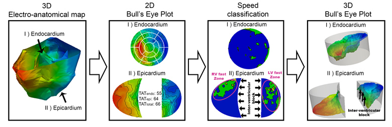
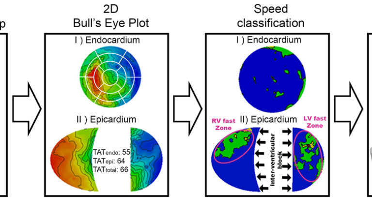
Electro-anatomical maps (EAMs) are commonly acquired in clinical routine for guiding ablation therapies. They provide voltage and activation time information on a 3D anatomical mesh representation, making them useful for analyzing the electrical activation patterns in specific pathologies. However, the variability between the different acquisitions and anatomies hampers the comparison between different maps. This paper presents two contributions for the analysis of electrical patterns in EAM data from biventricular surfaces of cardiac chambers. The first contribution is an integrated automatic 2D disk representation (2D BEP) of the LV and RV obtained with a quasi-conformal mapping from the 3D EAM meshes, that allows an analysis of Cardiac Resynchronization Therapy (CRT) lead positioning, interpretation of global (total activation time) and local indices (local activation time, surrogates of conduction velocity, inter-ventricular and transmural delays) that characterize changes in the electrical activation pattern. The second contribution is a set of indices derived from the electrical activation: speed maps, computed from LAT values, to study the electrical wave propagation, and histograms of isochrones to analyze regional electrical heterogeneities in the ventricles. We have applied the proposed methods to look for the underlying physiological mechanisms of LBBB and CRT, with the goal of optimizing the therapy by improving CRT response. To better illustrate the benefits of the proposed tools, we created a set of synthetically generated and fully-controlled activation patterns, where the proposed representation and indices were validated. Then, the proposed analysis tools are used to analyze EAM data from an experimental swine model of induced Left Bundle Branch Block (LBBB) with an implanted CRT device. We have analyzed and compared electrical activation patterns at baseline, LBBB and CRT stages in four animals: two without any structural disease and two with an induced infarction. By relating the CRT lead location with electrical dyssynchrony, we evaluated current hypotheses about lead placement in CRT and showed that optimal pacing sites should target the RV lead close to the apex and the LV one distant from it.
This study was partially funded by the Spanish Ministry of Science and Innovation (TIN2011-28067), the Spanish Industrial and Technological Development Center (cvREMOD-CEN-20091044), the Seventh Framework Programme (FP7/2007-2013) for research, technological and demonstration under grant agreement VP2HF (nº 611823), and the Spanish Ministry of Economy and Competitiveness under the Maria de Maeztu Units of Excellence Programme (MDM-2015-0502).
Quantitative Analysis of Electro-Anatomical Maps: Application to an Experimental Model of LBBB/CRT
Sign-in or become an IEEE member to discover the full contents of the paper.