

Abstract
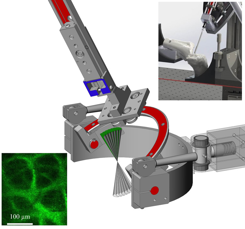
Previous studies using nonlinear microscopy have demonstrated that osteoarthritis (OA) is characterized by the gradual replacement of Type II collagen with Type I collagen. The objective of this study was to develop a prototype nonlinear laser scanning microendoscope capable of resolving the structural differences of collagen in various orthopaedically relevant cartilaginous surfaces. The current prototype developed a miniaturized femtosecond laser scanning instrument, mounted on an articulated positioning system, capable of both conventional arthroscopy and second-harmonic laser-scanning microscopy. Its optical system includes a multi-resolution optical system using a gradient index objective lens and a customized multi-purpose fiber optic sheath to maximize the collection of backscattered photons or provide joint capsule illumination. The stability and suitability of the prototype arthroscope to approach and image cartilage were evaluated through preliminary testing on fresh, minimally processed, and partially intact porcine knee joints. Image quality was sufficient to distinguish between hyaline cartilage and fibrocartilage through unique Type I and Type II collagen-specific characteristics. Imaging the meniscus revealed that the system was able to visualize differences in the collagen arrangement between the superficial and lamellar layers. Such detailed in vivo imaging of the cartilage surfaces could obviate the need to perform biopsies for ex vivo histological analysis in the future, and provide an alternative to conventional external imaging to characterize and diagnose progressive and degenerative cartilage diseases such as OA. Moreover, this system is readily customizable and may provide a suitable and modular platform for developing additional tools utilizing femtosecond lasers for tissue cutting within the familiar confines of two or three portal arthroscopy techniques.