
This Article is Featured in the Special Issue NIH-IEEE POCT 2015
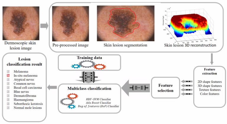
Melanoma mortality rates are the highest amongst skin cancer patients. Melanoma is life-threating when it grows beyond the dermis of the skin. Hence, depth is an important factor to diagnose melanoma. This paper introduces a non-invasive computerized dermoscopy system that considers the estimated depth of skin lesions for diagnosis. A 3-D skin lesion reconstruction technique using the estimated depth obtained from regular dermoscopic images is presented. On basis of the 3-D reconstruction, depth and 3-D shape features are extracted. In addition to 3-D features, regular color, texture, and 2-D shape features are also extracted. Feature extraction is critical to achieve accurate results. Apart from melanoma, in-situ melanoma the proposed system is designed to diagnose basal cell carcinoma, blue nevus, dermatofibroma, haemangioma, seborrhoeic keratosis, and normal mole lesions. For experimental evaluations, the PH2, ISIC: Melanoma Project, and ATLAS dermoscopy data sets is considered. Different feature set combinations is considered and performance is evaluated. Significant performance improvement is reported the post inclusion of estimated depth and 3-D features. The good classification scores of sensitivity = 96%, specificity = 97% on PH2 data set and sensitivity = 98%, specificity = 99% on the ATLAS data set is achieved. Experiments conducted to estimate tumor depth from 3-D lesion reconstruction is presented. Experimental results achieved prove that the proposed computerized dermoscopy system is efficient and can be used to diagnose varied skin lesion dermoscopy images.