
Abstract

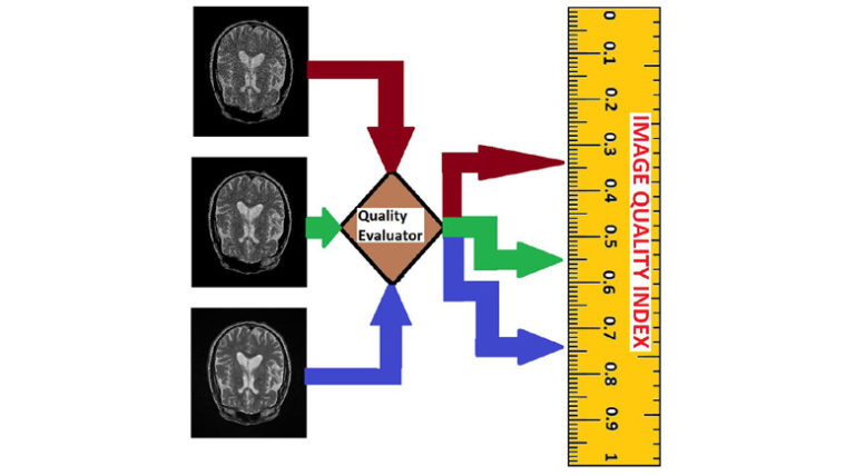
MRI system images are important components in the development of drugs because it can reveal the underlying pathology in diseases. Unfortunately the processes of image acquisition, storage, transmission, processing and analysis can influence image quality with the risk of compromising the reliability of MRI-based data. Therefore, it is necessary to monitor image quality throughout the different stages of the imaging workflow. This report describes a new approach to evaluate the quality of an MRI slice in multi-center clinical trials. The design philosophy assumes that an MRI slice, like all natural images, possess statistical properties that can describe different levels of contrast degradation. A unique set of pixel configuration is assigned to each possible level of contrast-distorted MRI slice. Invocation of the central limit theorem results in two separate Gaussian distributions. The central limit theorem says that the mean and standard deviation of pixel configuration assigned to each possible level of contrast degradation will follow a normal distribution. The mean of each normal distribution corresponds to the mean and standard deviation of the underlying ideal image. Quality prediction processes for a test image can be summarized into four steps. The first step extract local contrast feature image from the test image. The second step computes the mean and standard deviation of the feature image. The third step separately standardizes each normal distribution using the mean and standard deviation computed from the feature image. This gives two separate z-scores. The fourth step predicts the lightness contrast quality score and the texture contrast quality score from cumulative distribution function of the appropriate normal distribution. The proposed method was evaluated objectively on brain and cardiac MRI volume data using four different types and levels of degradation. The four types of degradation are Rician noise, circular blur, motion blur and intensity nonuniformity also known as bias fields. Objective evaluation was validated using a proposed variation of difference of mean opinion scores. Results from performance evaluation show that the proposed method will be suitable to monitor and standardize image quality throughout the different stages of imaging workflow in large clinical trials. Matlab implementation of the proposed objective quality evaluation method can be downloaded from (https://github.com/ezimic/Image-Quality-Evaluation).