
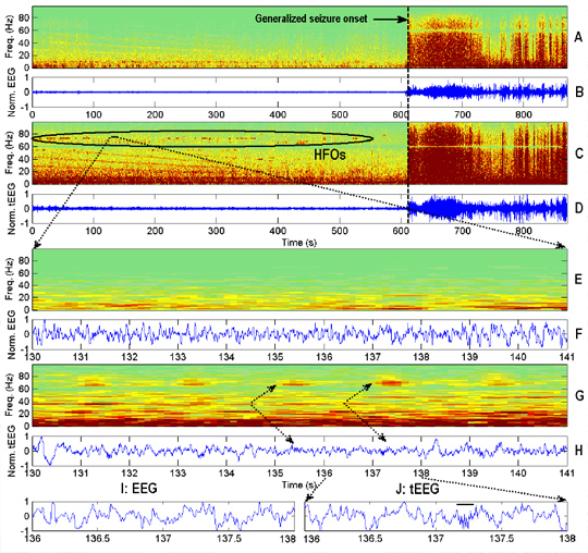

from Panel B at the onset of the generalized seizure (dashed line). Panels C, D, and F are the corresponding tEEG signals from Fp2’ (1-100 Hz, 200 S/s). Note
the high gamma-band burst HFOs just prior to the partial seizure (highlighted by ellipse in panel C).
Epilepsy is the second most prevalent neurological disorder (~1% prevalence) affecting approximately 67 million people worldwide with up to 75% from developing countries. The conventional electroencephalogram is plagued with artifacts from movements, muscles, and other sources. Tripolar concentric ring electrodes automatically attenuate muscle artifacts and provide improved signal quality. We performed basic experiments in healthy humans to show that tripolar concentric ring electrodes can indeed record the physiological alpha waves while eyes are closed. We then conducted concurrent recordings with conventional disc electrodes and tripolar concentric ring electrodes from patients with epilepsy. We found that we could detect high frequency oscillations, a marker for early seizure development and epileptogenic zone, on the scalp surface that appeared to become more narrow-band just prior to seizures. High frequency oscillations preceding seizures were present in an average of 35.5% of tripolar concentric ring electrode data channels for all the patients with epilepsy whose seizures were recorded and absent in the corresponding conventional disc electrode data. An average of 78.2% of channels that contained high frequency oscillations were within the seizure onset or irritative zones determined independently by three epileptologists based on conventional disc electrode data and videos.
View full article