Early Access Note:
Early Access articles are new content made available in advance of the final electronic or print versions and result from IEEE’s Preprint or Rapid Post processes. Preprint articles are peer-reviewed but not fully edited. Rapid Post articles are peer-reviewed and edited but not paginated. Both these types of Early Access articles are fully citable from the moment they appear in IEEE Xplore.

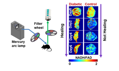
Background: Diabetes is known to cause delayed wound healing, and chronic non-healing lower extremity ulcers may end with lower limb amputations and mortalities. Given the increasing prevalence of diabetes mellitus worldwide, it is critical to focus on underlying mechanisms of these debilitating wounds to find novel therapeutic strategies and thereby improve patient outcome. Methods: This study aims to design a label-free optical fluorescence imager that captures metabolic indices (NADH and FAD autofluorescence) and monitors the in vivo wound healing progress noninvasively. Furthermore, 3D optical cryo-imaging of the mitochondrial redox state was utilized to assess the volumetric redox state of the wound tissue. Results: The results from our in vivo fluorescence imager and the 3D cryo-imager quantify the differences between the redox state of wounds on diabetic mice in comparison with the control mice. These metabolic changes are associated with mitochondrial dysfunction and higher oxidative stress in diabetic wounds. A significant correlation was observed between the redox state and the area of the wounds. Conclusion: The results suggest that our developed novel optical imaging system can successfully be used as an optical indicator of the complex wound healing process noninvasively.