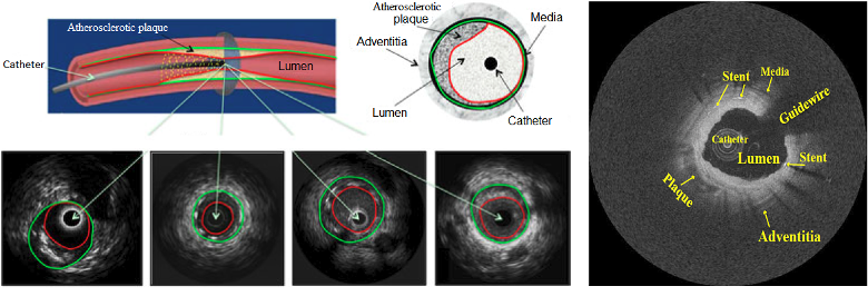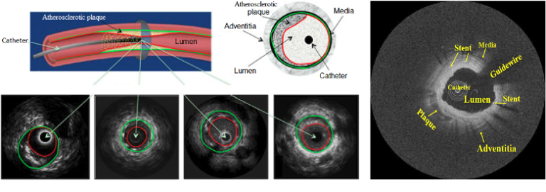
Minimally invasive transcatheter technologies have demonstrated substantial promise for the diagnosis and the treatment of cardiovascular diseases. For example, transcatheter aortic valve implantation is an alternative to aortic valve replacement for the treatment of severe aortic stenosis, and transcatheter atrial fibrillation ablation is widely used for the treatment and the cure of atrial fibrillation. In addition, catheter-based intravascular ultrasound and optical coherence tomography imaging of coronary arteries provides important information about the coronary lumen, wall, and plaque characteristics. Qualitative and quantitative analysis of these cross-sectional image data will be beneficial to the evaluation and the treatment of coronary artery diseases such as atherosclerosis. In all the phases (preoperative, intraoperative, and postoperative) during the transcatheter intervention procedure, computer vision techniques (e.g., image segmentation and motion tracking) have been largely applied in the field to accomplish tasks like annulus measurement, valve selection, catheter placement control, and vessel centerline extraction. This provides beneficial guidance for the clinicians in surgical planning, disease diagnosis, and treatment assessment. In this paper, we present a systematical review on these state-of-the-art methods. We aim to give a comprehensive overview for researchers in the area of computer vision on the subject of transcatheter intervention. Research in medical computing is multi-disciplinary due to its nature, and hence, it is important to understand the application domain, clinical background, and imaging modality, so that methods and quantitative measurements derived from analyzing the imaging data are appropriate and meaningful. We thus provide an overview on the background information of the transcatheter intervention procedures, as well as a review of the computer vision techniques and methodologies applied in this area.

