
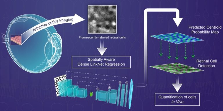
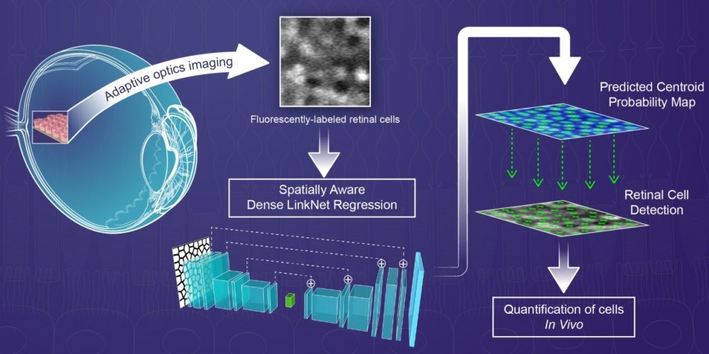
Recent advancements in ophthalmic imaging technology, based on adaptive optics (AO), have enabled fluorescence imaging of retinal pigment epithelial (RPE) cells directly inside the living human eye. RPE cells are affected in many blinding diseases but the hexagonally-packed RPE cell mosaic is difficult to visualize with traditional ophthalmic imaging tools which primarily provide tissue-level information. The improved resolution and contrast provided by AO fluorescence imaging gives rise to the possibility for automated RPE cell detection in AO images, an important step towards quantifying disease progression at the cellular level. This paper introduces a Spatially Aware Dense LinkNet Regression approach to detect RPE cells. The concept of spatial awareness, represented by Voronoi diagrams, is inspired by biology and based on the spatial arrangement of the RPE cell monolayer. Although cell centroid probability maps can be created by performing the distance transform on a set of manually-annotated cell centroids, here, the combination of spatially-aware constraint maps together with cell centroid probability maps improves RPE cell detection accuracy. To reduce model parameters in the dense-LinkNet regression, feature maps from the encode and decode paths are added. Automated detection of RPE cells determined from predicted centroid probability maps are used to evaluate cell density and spacing. Together, AO fluorescence imaging of RPE cells and automated detection of those cells enables quantitative assessment of cellular status in patient eyes.