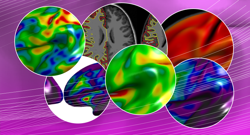
Brain anatomical MRI is increasingly viewed as an attractive way to measure biological properties of interest. In particular, numerous neuroimaging studies have shown that it is possible to distinguish patients from healthy control subjects using cortical thickness measurements. In this context, pooling data acquired on different MR scanners is a commonly used practice to increase the statistical power of studies based on MRI-derived measurements. Such studies are very appealing since they should make it possible to detect more subtle effects related to pathologies. However, the influence of confounds introduced by scanner-related variations remains unclear, it is thus crucial to investigate whether scanner-induced errors can exceed the effect of the disease itself. More specifically, in the context of developmental pathologies such as Autism Spectrum Disorders (ASD), it is essential to evaluate the influence of the scanner on age-related effects.
In this paper, we studied a dataset composed of 159 anatomical MR images pooled from three different scanners, including 75 ASD patients and 84 healthy controls. We quantitatively assessed the effects of the age, pathology and scanner factors on cortical thickness measurements. We observe that the effect size associated to the scanner factor is larger than the effect of pathology in almost every cortical region. Moreover, while the effect of age is consistent across scanners, the interaction between the age and scanner factors is important and significant in some specific cortical areas. Our results allow for a better understanding of the frequent discrepancies observed in the literature of ASD. We conclude that maximizing the consistency of image characteristics across scanners is mandatory for multi-site quantitative MRI studies in the context of neuro-developmental pathologies such as ASD.

