
S. Roychowdhury, D. D. Koozekanani, and K. K. Parhi

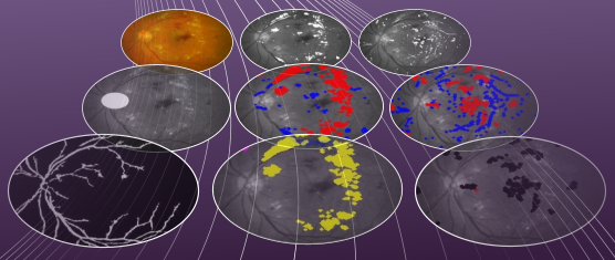
Diabetic retinopathy (DR) is the leading cause of blindness among people of working age in the developed world. Early screening and detection of DR can prevent about 90% of patients with early signs of DR from acquired blindness. Furthermore, automated prioritization of patients based on the severity of DR can increase the number of patients receiving timely eye-care by 50%. In such situations, automated screening programs using fundus images prior to manual grading can be extremely cost-effective and beneficial. This paper presents a three-stage automated fundus image analysis system called DREAM (Diabetic Retinopathy Analysis using Machine Learning) that detects the presence of mild, moderate and severe DR using fundus images. The first stage detects the blood vessels and optic disc region as the image background. Next, foreground candidate regions for bright and red lesions are extracted. In the second stage, foreground candidate regions for bright lesions are then classified as hard-exudates and cotton-wool spots, while the foreground candidate regions for red lesions are classified as hemorrhages and micro-aneurysms. Finally, in the third stage, the numbers of red lesions detected per image are combined to generate a DR severity grade. The proposed system assigns a grade of no DR, mild DR, moderate DR and severe DR, and achieves 100% sensitivity and 53% specificity for screening images with DR vs. healthy fundus images with no DR.
Tags: bright lesions, classification, diabetic retinopathy, fundus image processing, red lesions, segmentation, severity grade