Biomedical and health technology is progressing at breakneck speed. From specialty pharmacies to general discount shops, store shelves are packed with a vast assortment of wearable medical devices that measure glucose levels, heart rate, and other health metrics; and over-the-counter test kits are helping to check for a wide array of infections. At the same time, electronic health records and other data-sharing platforms have smoothed the mass shift from in-person to virtual office visits over the past two years, and new imaging technologies are allowing earlier disease detection so treatments can begin sooner when they are more effective.
These are only a few of the many advances that are already pushing the biomedical field into new territory. What other advances are primed to make their own mark on health care and biomedicine in the years to come? Here we highlight four technologies poised to make an impact as follows:
• three-dimensional (3D) printing of personalized medicines;
• the digital patient that acts as a person’s virtual twin;
• brain–machine interfaces to help patients with various neurological conditions;
• programmable, living robots for intelligent drug delivery or other areas.
Personalized medicine with 3D printing
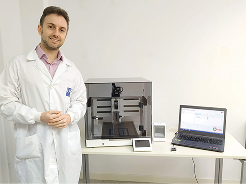
The 3D printing of medicines makes it possible for pharmacies and hospitals to produce medications on-demand and perfectly matched to the needs of individual patients. One of the companies pursuing this line of work is FabRx (pronounced like “fabrics”), a spinoff based on the work of University College London academics. Initially, the researchers hoped to adapt conventional printers, but were unable to find a printer company that was interested in retooling its hardware or software for pharmaceutical purposes. So, they did it themselves. “The technology is nothing new, but it needed the right design so it was easy to clean, could avoid contamination, and was especially suited for pharmaceutical products,” said Alvaro Goyanes, Ph.D., co-founder and director of development of FabRx Ltd., London, U.K. (Figure 1).

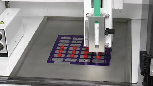
At the end of 2020, the company introduced its M3DIMAKER printer, which it promotes as the world’s first 3D printer for pharmaceuticals [1]. The printer is able to melt or crush drugs, and then use fused deposition modeling or direct powder extrusion to recombine them into 3D printed tablets, or “printlets.” It can produce a 28-printlet blister pack of made-to-order pills in about 10 minutes (Figure 2), Goyanes said. As an example of its utility, the company, in collaboration with Universidad de Santiago de Compostela, Santiago, Spain, tested the printer in a hospital. “We printed chewable formulations with different flavors for children who had rare metabolic diseases. These were formulations that weren’t available in the market,” he explained [2].

Currently, FabRx is participating in numerous clinical studies in humans and preclinical studies in animals to show M3DIMAKER’s potential for testing and developing new products, including printlets that combine more than one drug, Goyanes said. Along those lines, the company is working with the Paris hospital Gustave Roussy on blending a cancer drug with another medication that dampens its side effects. “We developed the formulations using 3D printing, and the clinical trial will start soon, hopefully to show that we can increase patient acceptability of the cancer drug,” he remarked. FabRx is also experimenting with adding raised braille symbols to tablets, which would be useful for visually impaired patients; or QR codes that could help nurses verify they are giving the right pill to the right patient, or link to online versions of patient information leaflets, he said.
Although he is sure that 3D-printed drugs are coming, Goyanes said it will likely take a few years. Since 3D-printing is an entirely new way of manufacturing medicines, it will need considerable clinical-trial data to win regulatory approval. It will also require the involvement of pharmaceutical companies, which he envisions will provide printer cartridges for their drugs so they can be easily inserted into the printer to produce on-demand, to-specification printlets. He remarked, “I don’t want to create high expectations that tomorrow everything is going to be 3D printed, but we will get there … even if it’s slowly.”
Digital patient
Digital patient, virtual twin, or health avatar: Whatever the terminology, this type of in silico model would integrate every little bit of medical-related data, including genetic and behavioral information, about an individual. This would raise the bar for personalized health care by allowing disease risks to be spotted, preventative measures to be taken, treatment options to be analyzed for their potential, and the effectiveness of selected treatments to be tracked over time. Such an all-encompassing digital patient is still a pipe dream, but researchers, like those at the Virtual Physiological Human Institute for Integrative Biomedical Research (VPH Institute), are taking steps in that direction.
An international nonprofit incorporated in Belgium in 2011, the VPH Institute now has more than 140 scientist members from 30 countries. Its mission is broad: to generate “individualized physiology-based computer simulations” for disease prevention, diagnosis, prognostic assessment, and treatment, as well as the development of biomedical products.
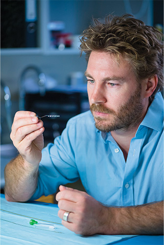
Acceleration of the drug-discovery pipeline is an area of special interest for VPH-affiliated researcher Francesco Pappalardo, Ph.D., professor of computer science at the Department of Drug and Health Sciences, University of Catania, Sicily, Italy (Figure 3). In particular, he and his research group are concentrating on employing a digital patient that includes clinical parameters ranging from immunological profiles to genetic analyses to “predict the way a specific drug is effective against a disease of interest, the optimal dosage, and the risks the drug can elicit on specific patient populations: elderly, pediatric, immune-depressed, and so on,” he said.

Toward that end, Pappalardo and his group have developed a computer modeling and simulation (M&S) platform that is able to simulate human immune-system dynamics, and have applied it to predict the effects of vaccines and other immunotherapies to a wide range of diseases, including tuberculosis, multiple sclerosis (MS), breast cancer, melanoma, influenza, and COVID-19. With COVID-19, for example, they predicted the partial inefficacy of hyperimmune plasma, the needs for monoclonal antibody-based therapies to be administered in the first phases of the disease, and the possibility that already-administered vaccines elicit cross-protection [3]. And for MS, they developed “a clinical decision-support system that could help medical doctors establish the best intervention based on the simulated digital patient,” he described [4].

The group is now improving on its M&S platforms for the drug-discovery pipeline to hasten a drug’s time to market, and as a result, hopefully reduce final price, which “could allow drugs to be distributed easily to low-income countries,” he said.
The possibilities are great, Pappalardo remarked. “Regulatory qualification of M&S platforms in health care is for sure the next step. It will allow health care to use M&S and digital patients in the same way traditional qualified methods are used now.”
Higher quality brain signals
The idea of a thought-controlled external device has drawn great interest, especially over the past two decades as researchers have developed new brain-to-machine interface (BMI) technologies for individuals who have visual, auditory, communication, or motor disabilities. Such technologies have primarily involved reading brain signals via electrodes placed on the scalp, or on the brain via craniotomy, but neither is perfect, explained Nick Opie, Ph.D., chief technology officer of the bioelectronics company Synchron Inc., which has offices in Melbourne, Australia, and Brooklyn, New York (Figure 4). “With scalp electrodes, the skull blocks the signal from the brain, so at least to date, they can’t acquire high-quality signals. Other technologies remove some of the skull and insert electrodes onto the surface of the brain or penetrate them directly into the brain itself, and while these have been shown to work very well and people have been able to use them to control (external machines), the surgery can cause damage to the brain, and there is also infection risk from having wires and leads coming out of your head,” he said.
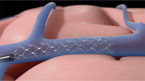
To solve both problems, Synchron is developing something different: a device that is inserted into the brain but with no need to cut a hole through the skull. Called Stentrode, the device is somewhat similar to a cardiac stent in that it is delivered through blood vessels to the target site, where it self-expands to conform to and press up against the vessel wall (Figure 5). Rather than simply opening the vessel, Opie said, the Stentrode has embedded electronics and wires capable of picking up clear brain signals, which then travel through a lead that exits the body to a telemetry/computer unit so they may be interpreted to operate various pieces of equipment.
An initial small trial of the Stentrode showed that people with paralysis could use it to control communication software and computers [5]. “These are patients who cannot move their arms, but with the device, they can use their mind to control a computer mouse,” Opie said. “They’ve loved that, and they’ve been really great working with us and telling us what other things they want to control: a smart home, messaging applications, and a whole range of different things.”
Currently, Synchron is expanding patient trials in Australia and is preparing to begin a clinical trial in the United States [6], he said. At the same time, company researchers are studying how they can use the technology to restore capabilities to patients with epilepsy, Parkinson’s disease, and a variety of other neurological conditions.

Of the biomedical potential of BMIs overall, Opie remarked, “The field is continually advancing; the technology is getting better and better; and the understanding of what’s needed both from the patient side and the technology side is also increasing to a level where I really do think that the paralysis problem will be overcome, and people will be able to have the lives they had before the injury or accident. It’s an exciting time, and I’m fortunate and thankful to be a part of it.”
Programmable, living robots
Programmable, living robots made their initial splash in 2020, and remain in the news today with the announcement that they are able to autonomously self-replicate (see “AI-Designed, Living Robots Can Self-Replicate” in this issue of IEEE Pulse). Called xenobots, they are made of skin cells collected from African clawed frogs, and directed into various configurations based on designs generated by an evolutionary algorithm. The idea was to explore the plasticity of cellular collectives – could genetically normal frog cells make something besides a tadpole?—and if so, to use machine learning to guide the self-assembly of cells into new structures with specific forms and functions.
Not only was it possible, but the research group, which comprises computer scientists and developmental biologists at Tufts and Harvard universities in Massachusetts and the University of Vermont, was able to make xenobots that could walk, swim, and gather inert materials into piles of certain sizes (Figure 6). In addition, the researchers showed that these xenobots could recover from injury, including being cleaved nearly in two, something far beyond the capability of even the most advanced machines constructed today.

These discoveries, plus the groundbreaking finding that the xenobots can self-replicate in a way that is completely different from any known form of biological reproduction, present myriad possibilities for biomedical research and health care advances [7], [8]. These include adding sensory capabilities to xenobots so they are able to assemble novel drugs or deliver drugs to specific sites in the human body for targeted action, or perhaps aid in environmental remediation, such as harvesting and removing microplastics from waterways, said team member Joshua Bongard, Ph.D., professor of computer science at the University of Vermont.
Team member Michael Levin, Ph.D., director of the Center for Regenerative and Developmental Biology at Tufts, is especially interested in xenobots’ applications in regenerative medicine, and the potential capability to eventually direct cells to do such things as develop new tissues to heal traumatic injuries, reprogram cancerous tissues, or even counteract aging. “If somebody has lost an arm, for example, you’re not going to recreate the arm directly from stem cells or through gene therapy because we have no idea yet how to micromanage that process at the level of individual cells or genes. But you might be able to communicate specific signals to the remaining cells to get them to regrow it the way that they did the first time around, but only if you understand something about how those cellular collectives make decisions like that,” he said. That is where xenobots come in. “This whole notion of learning to communicate with the collective and push it into the outcome that you want to see—beyond whatever the standard default course is—will eventually help us grow back organs and deal with all sorts of complex disease states.”
In addition to the wealth of potential applications, group member Douglas J. Blackiston, Ph.D., developmental biologist and senior scientist at the Tufts’ Allen Discovery Center, Harvard’s Wyss Institute, sees xenobots with a broader lens. “Everything that we think about in developmental biology and in regenerative medicine today is really aimed at understanding or rebuilding things that exist in nature,” he remarked. “Our work is about applying a bunch of varied techniques, and starting from scratch to create designer organisms for specific purposes just as we would build a (mechanical) robot or some other device.” he remarked.
“We are building something that has never existed in nature, could not be produced by natural selection, and is designed by a computer. And now we’re examining the ways we can get the system to replicate,” he added. “Every time we get a result, we’re left with so many more questions.”
References
- FabRx. Technologies. Accessed: Dec. 2, 2021. [Online]. Available: https://www.fabrx.co.uk/technologies/
- A. Goyanes et al., “Automated therapy preparation of isoleucine formulations using 3D printing for the treatment of MSUD: First single-centre, prospective, crossover study in patients,” Int. J. Pharmaceutics, vol. 567, Aug. 2019, Art. no. 118497.
- G. Russo et al., “In silico trial to test COVID-19 candidate vaccines: A case study with UISS platform,” BMC Bioinform., vol. 21, no. 17, p. 527, Dec. 2020.
- F. Pappalardo et al., “The potential of computational modeling to predict disease course and treatment response in patients with relapsing multiple sclerosis,” Cells, vol. 9, no. 3, p. 586, Mar. 2020.
- T. J. Oxley et al., “Motor neuroprosthesis implanted with neurointerventional surgery improves capacity for activities of daily living tasks in severe paralysis: First in-human experience,” J. Neurointervent. Surg., vol. 13, no. 2, pp. 102–108, Feb. 2021.
- University of Melbourne. (Jun. 8, 2021). Synchron Secures $52 Million to Launch U.S. Clinical Trials of Implantable Brain Device to Help Severely Paralysed Patients. Accessed: Dec. 5, 2021. [Online]. Available: https://about.unimelb.edu.au/news-resources/awards-and-achievements/announcements/synchron-secures-$52-million-to-launch-us-clinical-trials-of-implantable-brain-device-to-help-severely-paralysed-patients
- D. Blackiston et al., “A cellular platform for the development of synthetic living machines,” Sci. Robot., vol. 6, no. 52, p. eabf1571, Mar. 2021.
- S. Kriegman et al., “Kinematic self-replication in reconfigurable organisms,” Proc. Nat. Acad. Sci. USA, vol. 118, no. 49, Dec. 2021, Art. no. e2112672118.


